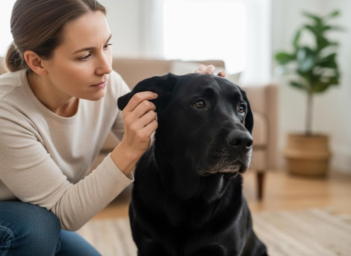
When your beloved pet experiences discomfort, especially with persistent ear issues, finding the root cause is paramount for their well-being. Traditional ear exams can provide valuable insights, but sometimes a more advanced approach is needed to truly understand the complexities of the ear canal. This is where video otoscopy shines, offering a detailed, magnified view that transforms diagnostic capabilities. At Great Plains Veterinary Dermatology, we believe in utilizing the most effective tools to provide clarity and the best possible care for your furry family members.
Unveiling the Depths: Why Video Otoscopy Goes Further
Many pet owners are familiar with the basic otoscope used during a routine vet clinic visit, which offers a limited, albeit helpful, view of the outer ear canal. However, chronic ear infections, deep-seated foreign bodies, or intricate growths often hide beyond this initial scope, making effective diagnosis a challenge. Video otoscopy takes this examination to an entirely new level. It employs a specialized, high-definition camera that can navigate the delicate and often convoluted pathways of your pet's ear canal, projecting a magnified image onto a screen. This allows for a much more thorough assessment, enabling a highly skilled vet dermatologist to identify issues that might otherwise be missed.
The Advanced Look: How Video Otoscopy Works
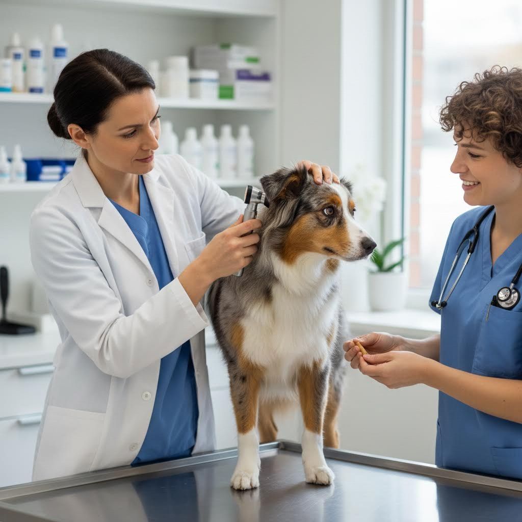
During a video otoscopy procedure, typically performed under sedation or general anesthesia for your pet’s comfort and safety, a small, flexible video endoscope is gently guided into the ear canal. This advanced tool allows the veterinarian to visualize the entire ear canal, including the eardrum, with unparalleled clarity. The high-resolution images and video footage are displayed on a monitor, allowing for precise identification of inflammation, polyps, tumors, foreign objects, or other abnormalities. Beyond diagnosis, this technology also facilitates targeted treatments, such as flushing stubborn debris, retrieving foreign bodies, or performing biopsies with precision, all while minimizing trauma to the sensitive ear structures.
A Collaborative Approach for Better Outcomes
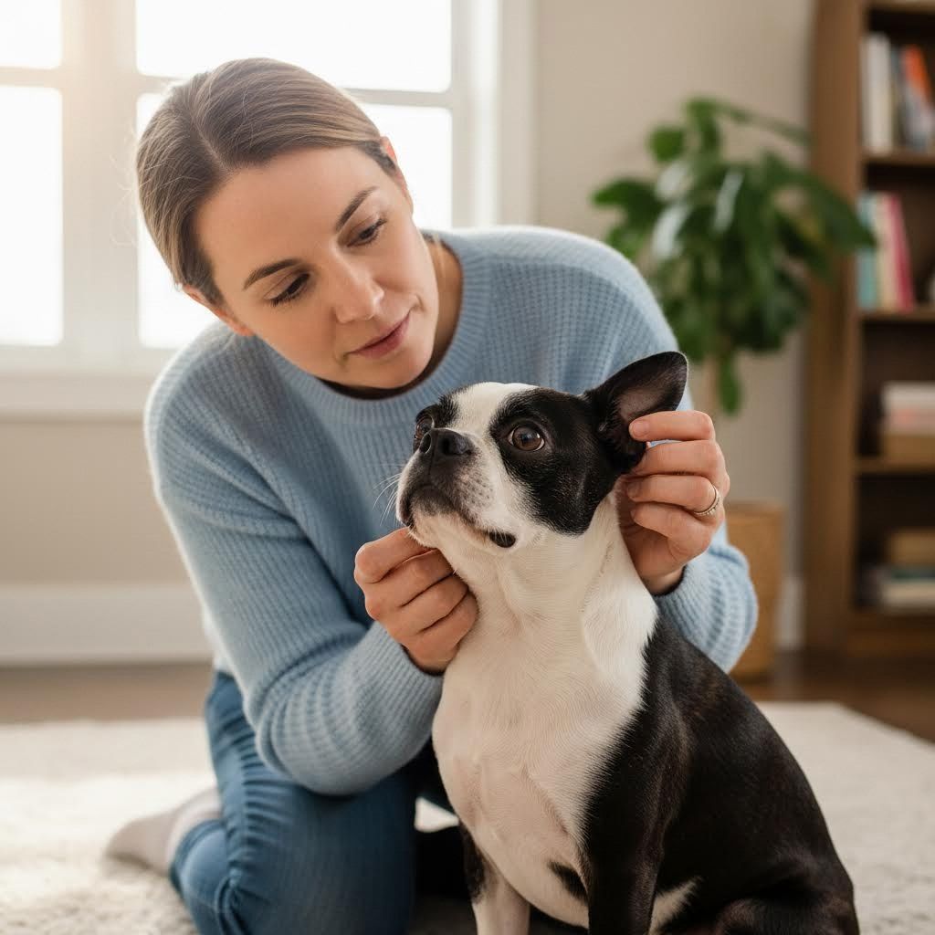
One of the most significant advantages of video otoscopy is the transparency it offers. Because the images are displayed on a screen, pet owners can often view the internal structures of their pet's ear canal alongside the board-certified veterinary dermatology specialist. This collaborative approach means you become an active partner in understanding your pet's condition and the rationale behind the recommended treatment plan. Seeing the problem firsthand fosters trust and helps reinforce the importance of adherence to follow-up care. This detailed visualization also enables a more accurate diagnosis, leading to more effective and sometimes less invasive treatments, ultimately saving your pet discomfort and potentially reducing long-term costs.
When your pet faces persistent or complex ear issues, understanding the problem fully is the first step toward lasting relief. Video otoscopy provides that clear understanding, offering an advanced diagnostic and therapeutic solution that goes far beyond traditional methods. At Great Plains Veterinary Dermatology, we are dedicated to employing cutting-edge techniques and our expertise in veterinary dermatology to ensure your pet receives the most precise and compassionate care. If you suspect your pet has a deeper ear issue, we encourage you to learn more about advanced ear examinations and how a specialist at our dermatology animal clinic in Omaha can help. Contact us today.